From Curiosity to Clinical Clarity: Teaching With a virtual dissection table That Learners Actually Love
November 01, 2025
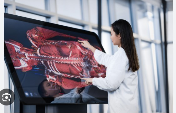
Anatomy clicks when structure, image, and implication meet in the same moment. A modern virtual dissection table makes that meeting routine: students rotate a life-size 3D body, peel layer by layer, slice in any plane, and jump to matching CT or MRI views without losing the thread. The effect is subtle but profound—less memorizing, more mapping; fewer disconnected facts, more navigable space. Below is a complete, classroom-ready playbook to turn your table into a dependable engine for pre-clinical mastery and clinical reasoning.
The value proposition in one sentence
A virtual dissection table converts passive look-and-label into active wayfinding: learners trace pathways, test hypotheses, and verify them instantly on cross-sectional imaging, all on a surface big enough for a team to think together.
What “usable realism” looks like
You don’t need every pixel on earth. You need fidelity that serves cognition:
- Layer control that respects true relationships. Skin → fascia → vessels → nerves → organ segments, with clean isolation and rapid fades so adjacency becomes obvious.
- Bidirectional plane sync. Slice the model axially, coronally, or sagittally and watch the equivalent imaging plane update; or start from a DICOM stack and project back to 3D.
- Multi-user touch that feels natural. Four to eight people can rotate, clip, measure, and pin labels at once without fighting the interface.
- One-tap “state save.” Faculty capture a view with labels and use it next week as an OSCE station—no remakes.
If those four boxes are ticked, everything else is a bonus.
A 10-week arc that compacts time to understanding
Replace one giant anatomy event with a weekly rhythm that compounds; keep sessions 60–75 minutes, prep under 15.
Week 1 — Planes, anchors, and orientation speed
Teach axial/coronal/sagittal with three anchors (heart, liver, knee). Drill: “Stabilize plane, find two landmarks, and rotate to improve the view.”
Week 2 — Vessels as the road network
Walk portal vs systemic pathways; show where mixing happens. Drill: “Trace a drop of contrast from splenic vein to right atrium.”
Week 3 — Nerve courses and danger zones
Map brachial plexus, facial nerve, sciatic pathway; overlay common entrapments. Case bite: “Foot drop after hip surgery—pin the most likely injury site.”
Week 4 — Segmental logic for surgeons
Hepatic segments, bronchopulmonary segments, renal vascular segments; draw safe resection lines and defend them on cross-sections.
Week 5 — Imaging literacy by doing
Sync the table with CT abdomen and MRI brain. Drill: “Find it on 3D, prove it on axial, then show one coronal slice that clarifies the relationship.”
Week 6 — Routes of spread
Air, blood, pus, and tumor: peritoneal reflections, retroperitoneal tracks, sinus pathways. Mini-scenarios contrast appendiceal perforation vs subphrenic abscess.
Week 7 — Procedures and safe windows
Central lines, chest tubes, lumbar puncture, abdominal incisions; mark surface landmarks, then verify the path on slices.
Week 8 — Variants that change decisions
Aberrant right subclavian, replaced hepatic arteries, renal accessory vessels, Circle of Willis variants—identify, risk, workaround.
Week 9 — Neuro navigation
Internal capsule, thalamic nuclei, brainstem sections; correlate named syndromes with lesions in two planes.
Week 10 — OSCE rehearsal and portfolio export
Timed stations mixing identify/relate/orient/explain. Auto-save results and prescribe 5-minute micro-reps for weak categories.
Teach relationships, not lists
Structure names are brittle; relationships are durable. Bake these prompts into every task:
- “Deep to this?” Forces true 3D reasoning.
- “Neighbor at risk?” Trains surgical caution.
- “Rotate to improve.” Builds plane literacy.
- “If injured, expect…?” Links structure to symptom in one line.
When questions demand relationships, recall becomes reasoning.
A scoring spine that motivates, not intimidates
Keep assessment transparent and fast:
- Locate (2 pts): tap or outline within a tolerance zone.
- Relate (2 pts): name the closest high-risk neighbor or “what lies deep to this plane.”
- Orient (1 pt): state current plane and the rotation to improve visibility.
- Explain (2 pts): one-sentence clinical implication.
- Time bonus (up to 3): finish early without hints.
Show scores instantly; tag misses by category (“ureter vs uterine artery,” “MCA vs ACA territory”) so next week’s micro-rep is obvious.
Turn imaging into a first-language skill
Don’t silo radiology until clerkships. Embed it from day one:
- Lock the model to slices. Walk a vessel or duct level by level; watch it morph on 3D.
- Flip mid-task. “Great—now find that on coronal.” Mental rotation becomes automatic.
- Add ultrasound windows. Overlay probe icons and typical views; reconcile surface landmarks with sonography.
Orientation lag evaporates when learners pivot across views a dozen times per session.
Five “tomorrow-ready” drills
- Stroke corridors: outline MCA/ACA/PCA territories on 3D; confirm on axial MRI; predict two deficits from two lesions.
- Pelvic thread-through: trace the ureter kidney→bladder, stop at three danger points, and show safe retraction paths.
- Chest tube confidence: place a tube on the surface model; verify on coronal CT that it tracks the safe triangle.
- Portal puzzles: follow portal triads; sketch a segment-sparing liver resection line; justify on imaging.
- Knee anchors: show ACL/PCL footprints; correlate with sagittal MRI; link to drawer tests.
Each ends with a 2-minute “find-it-fast” race for scan speed.
Faculty workflow that saves hours
If prep takes a morning, it won’t scale. Aim for:
- Reusable playlists (Thorax 2, Pelvis 1, Acute Abdomen) with saved “states” and hidden labels.
- Two-button capture to convert today’s view into an OSCE station.
- Lead-and-follow mode so one instructor guides multiple tables in sync.
- One-tap reset between cohorts.
Time saved becomes time taught.
Inclusive by design
Raise the baseline by making sessions accessible:
- High-contrast label modes and color-blind-friendly palettes.
- Captions on saved walkthroughs; optional stylus and large-target UI.
- Mirrored text prompts on a side display for learners who process better via words.
Inclusion isn’t extra credit—it’s how you lift the whole cohort.
Governance and data hygiene you can live with
If you import real studies:
- De-identify before load; simple approval flow for non-library cases.
- Role-based access (faculty edit, students annotate); audit of changes.
- Versioned “gold master” playlists so lessons never get overwritten.
A little hygiene keeps IT happy and classes uninterrupted.
Room setup that multiplies clarity
Treat the table like a shared microscope:
- Glare control (matte walls, adjustable lighting).
- 360° access with a low-profile ring so groups can approach from any side.
- Mirrored monitor for bigger rooms; acoustic damping for clean instructor audio.
Small environmental choices create big cognitive wins.
Measure what matters (and ignore the rest)
Lean analytics keep you focused:
- Orientation time: seconds to steady plane + anchor two landmarks.
- First-try accuracy: correct taps without hints.
- Transfer delta: gap between 3D ID and CT/MRI ID of the same structure.
- Two-week retention: error drop on repeat mini-quizzes.
- Throughput: learners served per hour without congestion.
Use a one-page dashboard to tune playlists, not to rank people.
Maintenance = trust
Uptime is pedagogy:
- Quarterly touch and color calibration; keep spare styluses and screen wipes at elbow height.
- “Clean slate” button for guaranteed resets.
- One-page rescue card for common hiccups; log fixes so trends surface early.
When the table never flakes, it earns a permanent slot on the schedule.
Extending beyond pre-clinicals
One platform, many departments:
- Surgery: incision planning, segmental resections, nerve-sparing corridors, trocar mapping.
- Emergency medicine: trauma windows, pneumothorax landmarks, fast stroke orientation.
- OB/GYN: pelvic floor mechanics, uterine blood supply, ureter safety along the uterine artery.
- Neurology/Neurosurgery: internal capsule, deep nuclei, brainstem cross-sections tied to named syndromes.
- Anesthesia & Pain: airway geometry, epidural routes, plexus blocks with US correlation.
- Physiotherapy/Sports: attachments, myotomes, kinematics linked to exam maneuvers.
Shared mental models across departments accelerate real-world collaboration.